
In What Causes Acne? and Why Do Pores Get Clogged?, we kept mentioning the same structure without stopping to explain it properly: the follicle.
It’s time to slow down and actually look at it.
Much of what we’ve discussed so far — healthy shedding, clogged pores and microcomedones — happens within one small structure: the pilosebaceous unit. Understanding what it is and how its parts work together makes the rest of the acne story much easier to follow.
What is the pilosebaceous unit?
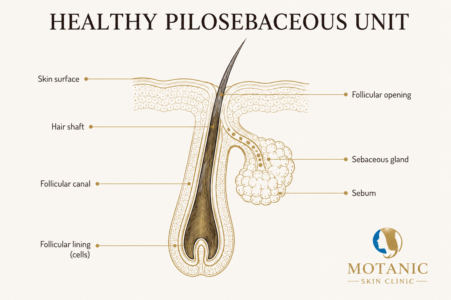
The pilosebaceous unit is the hair follicle together with its associated sebaceous gland.1 It’s a small structure, but many of the changes involved in acne take place within it — not on the surface of your skin, but just beneath it.
The pilosebaceous unit also includes a tiny arrector pili muscle attached to the follicle. Its main role relates to hair movement rather than the acne processes we’re focusing on here, so we won’t explore it further in this article.1
Starting at the surface: the follicular opening
What you see on the skin’s surface — what people commonly call a pore in this context — is the follicular opening. It’s the entrance to the follicle that extends beneath the skin.
The follicular canal
Just beneath the opening is the follicular canal — a narrow passage that extends down into the skin. The hair shaft travels upward through this canal and emerges at the surface. In acne-prone follicles, this canal is also where some of the earliest changes we described in Why Do Pores Get Clogged? take place. In a healthy follicle, cells lining the canal are continually renewed and shed without accumulating into a plug.
The hair shaft
The hair shaft travels upward through the follicular canal and emerges at the skin’s surface. In many acne-prone areas, the hair can be extremely fine and barely visible. For the purposes of understanding acne, the hair itself isn’t the important part — what matters is the canal it travels through, and what’s happening around it.
The sebaceous gland and sebum
Attached to the follicle is a small sebaceous gland. It opens onto the follicle through a short duct and produces sebum, an oily substance that helps protect and lubricate the skin.1
Sebum is produced through a process called holocrine secretion — the gland’s cells break down and release their contents as sebum, rather than simply excreting a fluid the way many other glands do.1 Sebaceous gland activity is also sensitive to hormones: androgens in particular stimulate the gland to produce more sebum.123
The follicular lining
The follicular canal is lined with cells called keratinocytes — the same broad category of cell found throughout the outer layers of your skin. Under normal conditions, these cells mature, move toward the surface, and shed in an orderly, continuous process. We won’t go into the cellular detail of how they work here; that’s its own subject.
Every part of this structure has an ordinary job. Acne develops when normal processes within this system begin to change.
How the system normally works together
In a healthy pilosebaceous unit, these processes tend to run quietly and continuously: the sebaceous gland produces sebum, while cells lining the follicular canal mature and shed in an orderly way. Sebum moves through the follicular canal toward the skin’s surface, while cells lining the canal are continually renewed and shed. This is close to the “before” picture we described in What Causes Acne? — the baseline that the early changes in acne development move away from.
What’s Really Happening Inside Your Skin?
See the healthy pilosebaceous unit explained visually, before we get into what changes when acne develops.
Why this structure matters for acne
Much of what we’ve discussed so far — altered shedding, sebum, the earliest plug formation we call a microcomedone — takes place within the pilosebaceous unit, beneath the surface of the skin, before anything becomes visible.
That’s really the point of this article: once you can picture the structure itself, the rest of the acne story has somewhere to happen.
References
- Martel JL, Miao JH, Badri T, Fakoya AO. Anatomy, Hair Follicle. StatPearls [Internet]. NCBI Bookshelf NBK470321.
- Sutaria AH, Masood S, Saleh HM, Schlessinger J. Acne Vulgaris. StatPearls [Internet]. NCBI Bookshelf NBK459173.
- Toyoda M, Morohashi M. Pathogenesis of acne. Medical Electron Microscopy. 2001;34(1):29–40.