

Cutibacterium acnes has acne in its name. It lives in the follicles where acne develops. And it can interact with the immune system in ways that contribute to inflammation.
So the conclusion seems obvious: bacteria causes acne.
Except there is a problem with that explanation.
C. acnes also lives on healthy skin.
It is a normal member of the skin microbiome and is particularly abundant in sebum-rich areas such as the face and back.1 Its presence alone therefore cannot explain why acne develops.
Instead of asking only whether C. acnes is present, we need to ask a more informative question:
What is happening in the follicle in which that organism is living?
What exactly is Cutibacterium acnes?
C. acnes is a bacterium strongly associated with human skin and particularly abundant in sebaceous regions. It is well adapted to the environment of the pilosebaceous unit — the same anatomical structure we explored in Understanding the Pilosebaceous Unit.1
If you’ve read older acne literature, you may recognise another name: Propionibacterium acnes, or P. acnes. That isn’t a different acne bacterium. Taxonomic work led to the organism being placed in the genus Cutibacterium, giving us the name Cutibacterium acnes.2
But the terminology is less important than the biological point: C. acnes is not simply a foreign organism that appears when someone develops acne. It is also found on healthy skin.1
If C. acnes lives on healthy skin, why doesn’t everyone develop acne?
This is where the familiar phrase “acne-causing bacteria” becomes too simplistic.
The role of C. acnes cannot be explained simply by whether the organism is present or absent. Research increasingly examines differences in microbial community structure, C. acnes phylotype composition and host–microbe interactions when trying to understand its role in acne.34
C. acnes is a species, but populations within that species are not genetically identical. Different phylogenetic groups — or phylotypes — can coexist on human skin, and their relative distribution has become an important area of acne research.3
Presence is not the same thing as causation.
That doesn’t mean C. acnes is irrelevant to acne. It means that “Is the bacterium there?” is not enough to explain what is happening.
The follicle around the bacterium matters
A microorganism inside a follicle does not exist in isolation.
It exists within a biological environment containing sebum, follicular cells, other microorganisms and host immune systems. Throughout this Knowledge Library, we’ve already examined several interconnected changes that can occur in acne-prone follicles.
In What Is Follicular Hyperkeratinisation?, we examined altered follicular shedding and retention.
In Sebum and Acne, we looked at the role of sebum without reducing acne to simply “oily skin.”
In What Is a Comedone?, we followed the development of microcomedones and visible comedonal lesions.
And in What Causes Inflammation in Acne?, we examined why visible redness is not necessarily the moment inflammatory biology begins.
C. acnes exists within this wider system. Reviews of acne and the cutaneous microbiome describe interactions between microbial communities, skin physiology and host responses rather than reducing the disease to one microorganism operating independently.45
Where Does C. acnes Fit Into the Acne Story?
Cutibacterium acnes naturally lives on human skin — so why does acne develop in some follicles? In Episode 8 of Rethinking Acne, we look inside the follicle to understand how its changing environment can influence the role of C. acnes, and why simply having the bacterium on your skin does not automatically mean you have acne.
Rethinking Acne · Episode 8 — understanding C. acnes as part of the wider follicular environment.
Not every C. acnes population is identical
Researchers classify C. acnes into different phylogenetic groups, or phylotypes, with additional variation at strain level.
Studies have reported differences in C. acnes population structure between acne-associated and healthy skin. Reduced phylotype diversity and greater representation of particular type I lineages, including IA1, have been reported in acne-associated populations.36
But this needs careful interpretation.
IA1 should not simply be labelled “the bad acne bacterium.”
Experimental research has shown that different phylotype contexts can produce different inflammatory responses. For example, one skin-explant study reported greater upregulation of several innate immune markers when skin was exposed to IA1 alone than when IA1 was combined with phylotypes II and III.6 That supports the importance of population context, but it does not establish a universal good-versus-bad classification for every strain or every person.
A 2026 study provides another useful example. Researchers examined skin swabs from 14 people with acne and 14 healthy participants. Healthy samples contained nearly equal quantities of phylotype IA and phylotypes IB/II, whereas acne samples showed a strong predominance of IA over IB/II. Importantly, that difference was driven mainly by marked depletion of IB/II rather than an increase in IA.7
Because this was a small study, its findings should not be treated as a universal rule for every person with acne. What it demonstrates particularly well is why a simple “more acne bacteria” explanation can miss what population-level measurements are actually showing.
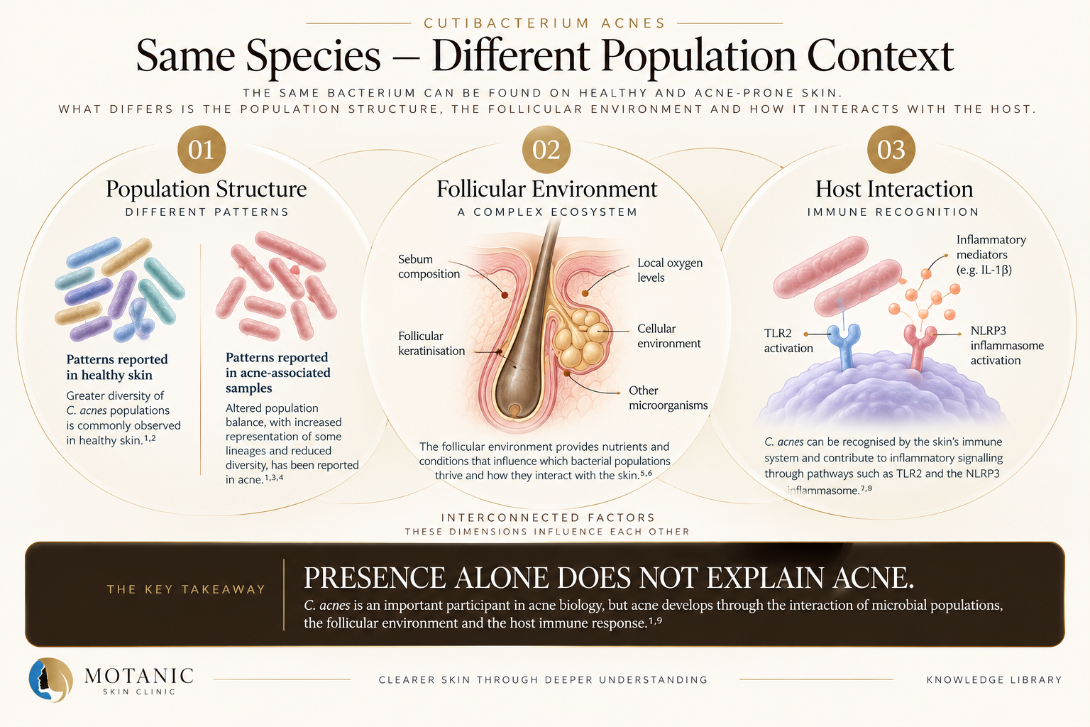
The presence of C. acnes alone does not explain acne. Population structure, the follicular environment and host immune interaction form part of the wider biological context.
The question may be less about whether C. acnes is present and more about which populations are present, in what balance, and in what follicular environment.
How can C. acnes contribute to inflammation?
If C. acnes normally lives on skin, how can it also participate in inflammatory acne?
Because normal resident does not mean biologically inactive.
Cells involved in the skin’s immune response possess mechanisms for recognising microbial signals. One important example is Toll-like receptor 2, or TLR2.
Experimental research has shown that C. acnes — then referred to as P. acnes — can activate inflammatory cytokine responses through TLR2. The same study identified TLR2-expressing macrophages around pilosebaceous follicles in acne lesions.8
Other innate immune mechanisms, including activation of the NLRP3 inflammasome and production of inflammatory mediators such as IL-1β, have also been investigated in acne biology.9
The important concept here isn’t memorising every receptor or cytokine. It is understanding the relationship:
Microbial signals can interact with host recognition systems and contribute to inflammatory signalling.
That’s the connection between this article and What Causes Inflammation in Acne?. Once again, the bacterium is participating within a biological system rather than acting independently of everything around it.
Is acne simply a bacterial infection?
No — that description does not adequately capture the biology.
People often hear the word bacteria and understandably think infection. But C. acnes is abundant on healthy, sebum-rich skin and contributes to normal skin ecology as well as being implicated in acne pathophysiology.1
Acne is described in the scientific literature as a chronic inflammatory disease with complex pathogenesis. Altered sebum production, inflammation, excess keratinisation and interactions involving the skin microbiome have all been implicated.35
That doesn’t make C. acnes unimportant.
It puts the bacterium in context.
So does C. acnes cause acne?
The evidence doesn’t fit neatly into a simple yes-or-no answer.
C. acnes is an important participant in acne pathophysiology. But simply having the bacterium on the skin is not sufficient to explain why acne develops: it is also found abundantly on healthy skin, while acne research has identified differences involving phylotype balance, microbial ecology and host inflammatory responses.134
So describing C. acnes simply as “the bacteria that causes acne” leaves out much of the biology.
What has changed in the follicular system that changes the relationship between the microorganism and its host?
That question connects C. acnes to everything we’ve already examined — follicular shedding, sebum, comedone formation and inflammation — instead of turning bacteria into the final isolated culprit.
Why this matters
The way we explain acne changes the way we think about it.
If acne is presented simply as bacteria, the intuitive response becomes: kill the bacteria.
If acne is presented simply as oil: remove the oil.
If acne is presented simply as blocked pores: unblock the pore.
Each explanation takes one component of acne biology and turns it into the whole disease.
The evidence we’ve followed throughout this series points toward something more interconnected.
The pilosebaceous unit provides the anatomical setting. Altered follicular keratinisation affects how material is retained. Sebum contributes to the follicular environment. Comedones develop within that system. Immune signalling contributes to inflammation. And C. acnes participates within that environment.
Understanding the relationships between those processes is more informative than searching for one culprit.
We’ve now followed acne from the anatomy of the pilosebaceous unit through follicular retention, sebum, comedone formation, inflammation and the role of Cutibacterium acnes. The next step in the Motanic Knowledge Library will build on that foundation rather than treating any one of these processes as the entire acne story.
Not sure what’s driving your acne?
Acne can involve several interacting factors, and what is happening can differ from one person to another. A skin consultation gives us the opportunity to assess your skin more closely and discuss an appropriate approach for your individual concerns.
Book a Skin ConsultationReferences
- Rozas M, Hart de Ruijter A, Fabrega MJ, et al. From Dysbiosis to Healthy Skin: Major Contributions of Cutibacterium acnes to Skin Homeostasis. Microorganisms. 2021;9(3):628. doi:10.3390/microorganisms9030628.
- Scholz CFP, Kilian M. The natural history of cutaneous propionibacteria, and reclassification of selected species within the genus Propionibacterium to the proposed novel genera Acidipropionibacterium, Cutibacterium and Pseudopropionibacterium. Int J Syst Evol Microbiol. 2016;66(11):4422–4432. doi:10.1099/ijsem.0.001367.
- Dréno B, Dekio I, Baldwin H, et al. Acne microbiome: From phyla to phylotypes. J Eur Acad Dermatol Venereol. 2024;38(4):657–664. doi:10.1111/jdv.19540.
- Dréno B, Dagnelie MA, Khammari A, Corvec S. The Skin Microbiome: A New Actor in Inflammatory Acne. Am J Clin Dermatol. 2020;21(Suppl 1):18–24. doi:10.1007/s40257-020-00531-1.
- Podwojniak A, Tan IJ, Sauer J, et al. Acne and the cutaneous microbiome: A systematic review of mechanisms and implications for treatments. J Eur Acad Dermatol Venereol. 2024. PMID: 39269130.
- Dagnelie MA, Corvec S, Saint-Jean M, Nguyen JM, Khammari A, Dréno B. Cutibacterium acnes phylotypes diversity loss: a trigger for skin inflammatory process. J Eur Acad Dermatol Venereol. 2019;33(12):2340–2348. doi:10.1111/jdv.15795.
- Feidenhansl C, Rruci E, Knödlseder N, Lomholt HB, Brüggemann H. Differential Quantification of Cutibacterium acnes Phylotypes IA and IB/II on Healthy and Acne-Prone Human Skin. Experimental Dermatology. 2026;35(8):e70347. PMID: 42603144.
- Kim J, Ochoa MT, Krutzik SR, et al. Activation of toll-like receptor 2 in acne triggers inflammatory cytokine responses. J Immunol. 2002;169(3):1535–1541. doi:10.4049/jimmunol.169.3.1535.
- Zhu W, Wang HL, Bu XL, Zhang JB, Lu YG. A narrative review of research progress on the role of NLRP3 inflammasome in acne vulgaris. Ann Transl Med. 2022;10(11):645. doi:10.21037/atm-21-5924.