
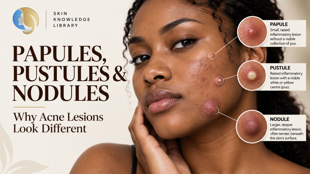
The different types of acne lesions can look surprisingly different from one another. Look closely at acne-prone skin and you may see a blackhead, a small skin-coloured bump, a raised inflammatory lesion, a spot with a visible white or yellow centre, or perhaps a deeper, tender lump beneath the surface.
In everyday conversation, all of these may simply be called pimples.
But biologically and clinically, they are not identical.
Acne can produce several different lesion types, including open and closed comedones, papules, pustules and deeper nodules. Importantly, several of these lesion types can occur on the same skin at the same time.
Understanding those differences helps us connect what we can see on the surface with what may be happening within individual follicles.
Acne lesions can share underlying follicular biology without every lesion following the same visible pathway.
Before the lesion has a name, start with the follicle
Although acne lesions can look very different from one another, they are centred on the pilosebaceous unit — the hair follicle and its associated sebaceous gland.
That matters because the visible lesion is only the surface expression of processes occurring within and around that follicular system.
Throughout the Motanic Skin Knowledge Library, we have been building that biology piece by piece.
We examined follicular hyperkeratinisation and abnormal retention within acne-prone follicles. We looked at sebum and acne, followed the development of microcomedones and comedones, explored inflammation in acne, and then placed Cutibacterium acnes within the wider follicular environment.
Now we can connect those processes with the lesions that become visible on the skin.
Research describes the microcomedone as an important subclinical precursor from which both inflammatory and non-inflammatory clinical acne lesions can emerge.
But there is an important qualification.
The older picture of acne as a perfectly ordered sequence — first a plug forms, then bacteria arrive, then inflammation begins — is too simple. Evidence indicates that inflammatory signalling can already be present during very early lesion development, including before or alongside microcomedone formation.
So we should not think of acne as a conveyor belt in which every follicle must pass through exactly the same visible stages.
Comedones: when there is no obvious inflammatory lesion
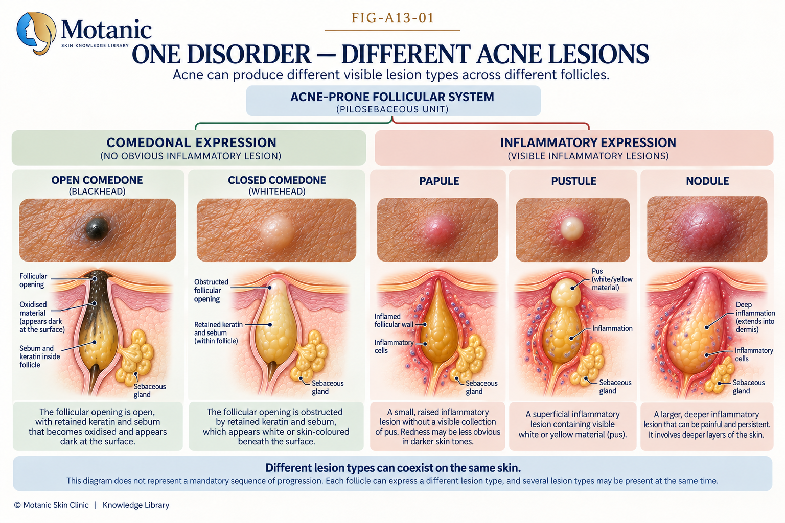
Two of the most familiar acne lesions are open comedones and closed comedones.
An open comedone is what we commonly call a blackhead. A closed comedone is what we commonly call a whitehead.
These are commonly classified as non-inflammatory acne lesions because they do not show the obvious clinical inflammatory features associated with papules and pustules.
That terminology should not be interpreted to mean that inflammatory biology is completely absent. Research into early acne development indicates that inflammatory activity can already be present during subclinical and comedonal stages.
We explored this process in much greater detail in What Is a Comedone? Blackheads, Whiteheads and Microcomedones Explained.
A comedone is one possible visible expression of an acne-prone follicular process — but it is not the only one.
What is an acne papule?
A papule is a small, raised inflammatory acne lesion without a visible collection of pus.
Papules are frequently described as red, tender bumps. But making redness part of the essential definition can create a problem: inflammation does not look identical on every skin tone.
In more deeply pigmented skin, erythema — the colour change associated with increased blood flow during inflammation — may be less visually obvious. The inflammatory lesion may therefore not present as the bright red bump often used to illustrate acne in lighter skin.
For that reason, describing the lesion by its morphology is more useful:
An acne papule is a small, raised inflammatory lesion without a visible collection of pus.
What is an acne pustule?
A pustule is another superficial inflammatory acne lesion, but unlike a papule it contains visible white or yellow purulent material.
This appearance can encourage a familiar chain of assumptions:
Pus → infection → bacteria → bacteria caused the acne.
But acne biology is more complicated than that.
Visible pus does not mean the skin is dirty, and its presence alone does not establish that acne is simply a bacterial infection.
As we explored in What Is Cutibacterium acnes — and Does Bacteria Really Cause Acne?, microorganisms are part of a wider follicular and inflammatory system. The presence of C. acnes alone is not sufficient to explain why acne develops.
A pustule is therefore better understood as one clinical expression of an inflammatory acne lesion rather than evidence that a “dirty pore” simply needs to be emptied.
What is an acne nodule?
Papules and pustules are relatively superficial inflammatory lesions.
A nodule represents deeper inflammatory involvement.
Clinically, acne nodules are larger, deeper inflammatory lesions and can be painful or tender.
This depth matters.
A small superficial papule and a deep painful nodule should not be treated as though they are merely different-sized versions of exactly the same visible bump. They represent different clinical morphologies and different depths of inflammatory involvement.
Deeper inflammatory acne is also important because acne can leave lasting changes, including scarring.
That does not mean every nodule will produce a scar. It means lesion depth and inflammatory severity are meaningful parts of the overall clinical picture.
What do people mean by “cystic acne”?
The term “cystic acne” is commonly encountered, but it can hide important differences between deep acne lesions.
Clinical descriptions may distinguish nodules from cyst-like or pseudocystic lesions. For that reason, not every deep, painful acne lesion should automatically be labelled a cyst.
For someone looking at their own skin, the goal is not to diagnose a nodule versus a pseudocyst in the mirror.
The more useful lesson is that a deep, painful inflammatory lesion is clinically different from a superficial blackhead, whitehead or small papule, and depth matters when acne is assessed.
Why can several acne lesion types appear at the same time?
This is one of the most important ideas in understanding the different types of acne lesions.
Imagine someone has several blackheads around the nose, closed comedones across the forehead, two inflammatory papules on one cheek, a pustule on the other cheek and one deeper tender nodule along the jawline.
It can look as though five completely different problems are happening.
But acne commonly contains several lesion types at the same time.
The better way to understand this is at the level of the individual follicle.
There are many pilosebaceous follicles across acne-prone areas of the face and body. They do not all have to be expressing the same visible morphology at the same moment.
One follicle may contain a microcomedone that cannot yet be easily seen. Another may present as a closed comedone. Another may have an open comedone. Another may show clinically apparent inflammatory activity as a papule or pustule. Another may be involved in a deeper inflammatory lesion.
Acne is occurring across many follicles. Different follicles can express different lesion morphologies at the same time.
This is why we should avoid teaching acne as:
whitehead → papule → pustule → nodule
as though every lesion must travel through those visible stages.
The microcomedone is an important precursor to subsequent clinical lesions, but inflammatory biology can begin early and acne lesion development is more complex than one mandatory visible sequence.
Can every acne lesion be extracted?
No. Seeing a raised or blocked-looking lesion does not automatically mean there is something that should be squeezed out.
Open comedones are the clearest comedonal targets for professional extraction when extraction is otherwise appropriate. Selected closed comedones may also be managed professionally, depending on the technique required, practitioner training, scope of practice and applicable infection-control requirements.
Papules, pustules and nodules are inflammatory lesions and should not simply be treated as blocked pores that need to be squeezed. A deeper inflammatory lesion is particularly different from a superficial comedone.
The important principle is simple: lesion visibility does not automatically mean lesion extractability.
Acne lesions in melanin-rich skin
The fundamental acne lesion types remain recognisable across skin tones, but their colour presentation and the changes that remain after a lesion resolves can differ.
This is particularly important in melanin-rich skin.
Post-inflammatory hyperpigmentation (PIH) can develop after inflammatory acne and is especially important in skin of colour. Reviews report greater prevalence, severity and persistence of acne-associated PIH in more deeply pigmented skin.
There is another important point.
Inflammation does not have to look dramatically red to have consequences.
In skin of colour, inflammation may sometimes be less visually obvious or more difficult to identify. Acne-associated PIH can also develop even where striking visible inflammation was not apparent.
That means relying only on bright redness as evidence of inflammation can give an incomplete picture, particularly when assessing melanin-rich skin.
The lesion can disappear before its consequences do
An inflammatory acne lesion may eventually flatten.
But that does not necessarily mean the skin immediately returns to how it looked before the lesion appeared.
Inflammation can be followed by post-inflammatory hyperpigmentation, leaving a flat area of increased pigmentation after the active lesion itself has resolved.
Acne can also leave textural scars, particularly in the context of more significant inflammatory disease.
This introduces an important distinction:
Active acne lesions and what acne leaves behind are related, but they are not the same thing.
A flat dark mark remaining after a pimple resolves is not necessarily active acne.
A depressed scar is not an active comedone.
And persistent colour change should not automatically be described as a scar.
Those distinctions deserve their own investigation.
Why lesion type matters
Learning the names of acne lesions is not about memorising dermatology vocabulary for its own sake.
Lesion morphology gives us information.
It helps distinguish predominantly comedonal presentations from clinically inflammatory ones. It helps describe whether lesions appear relatively superficial or deeper. It also allows the types and distribution of visible lesions to be documented more precisely.
But morphology is still only one part of acne assessment.
The types of lesions present, their number and distribution, the extent of involvement, and features such as pigmentary change or scarring can all contribute to the clinical picture.
That is why looking at one lesion and saying:
“That’s a pustule, therefore we know exactly why your acne is happening.”
would go beyond what lesion morphology alone can tell us.
The visible lesion is evidence. It is not the entire explanation.
The bigger acne picture
We have now travelled a long way beneath what most people simply call a pimple.
We started with the pilosebaceous unit.
We examined abnormal follicular retention and microcomedone formation.
We looked at sebum.
We explored comedones and inflammation.
We placed Cutibacterium acnes back into its wider biological context.
And now we can see how interconnected acne biology can produce different visible clinical expressions.
A blackhead, whitehead, papule, pustule and nodule do not need to represent five unrelated problems.
Nor should they be forced into one mandatory visible sequence.
What we see on the surface is the visible expression of processes occurring within individual follicles.
And sometimes the most important part of the story begins after the active lesion has gone.
That is where pigmentation and scarring enter the picture.
Continue Learning
The Motanic Skin Knowledge Library has now followed acne from follicular anatomy through microcomedone formation, sebum, inflammation, Cutibacterium acnes and the different lesions that become visible on the skin.
The next phase will examine what acne can leave behind — and why a flat dark mark, persistent colour change and a true acne scar should not automatically be treated as the same thing.
Not sure what type of acne you’re dealing with?
Acne can present with several lesion types at the same time, and the visible lesion is only one part of the overall picture. A skin consultation gives us the opportunity to assess the visible pattern of your skin alongside other relevant factors and discuss an appropriate approach for your individual concerns.
Book a Skin ConsultationReferences
- DermNet. Acne Vulgaris: Features, Types, and Treatments. Clinical overview of acne lesion morphology, pilosebaceous localisation, simultaneous lesion types and acne complications.
- Xie L, Hamblin MR, Zheng D, Wen X. The role of microcomedones in acne: Moving from a description to treatment target? J Dtsch Dermatol Ges. 2024;22(1):9–16. doi:10.1111/ddg.15272. PMID: 38123894.
- Del Rosso JQ, et al. The sequence of inflammation, relevant biomarkers, and the pathogenesis of acne vulgaris: what does recent research show and what does it mean to the clinician? J Drugs Dermatol. 2013. PMID: 23986176.
- Oulès B, Saurat JH. Strategic Targets in Acne, Update 2025: The Microcomedone Is Not Just a Plug, It Is an Egg. Dermatology. 2026;242(Suppl 1):8–14. doi:10.1159/000547515. PMID: 40924653.
- Shaheen B, Gonzalez M. Acne sans P. acnes. J Eur Acad Dermatol Venereol. 2012. PMID: 22458782.
- Chiang C, Ward M, Gooderham M. Dermatology: how to manage acne in skin of colour. Drugs Context. 2022;11:2021-10-9. doi:10.7573/dic.2021-10-9. PMID: 35720053.
- Davis EC, Callender VD. Postinflammatory Hyperpigmentation: A Review of the Epidemiology, Clinical Features, and Treatment Options in Skin of Color. J Clin Aesthet Dermatol. 2010;3(7):20–31.
- Callender VD, et al. Effects of Topical Retinoids on Acne and Post-inflammatory Hyperpigmentation in Patients with Skin of Color: A Clinical Review and Implications for Practice. Am J Clin Dermatol. 2022.
- Acne-induced Post-inflammatory Hyperpigmentation: From Grading to Treatment. Review of acne-associated PIH, including subtle inflammatory presentation and PIH in skin of colour.